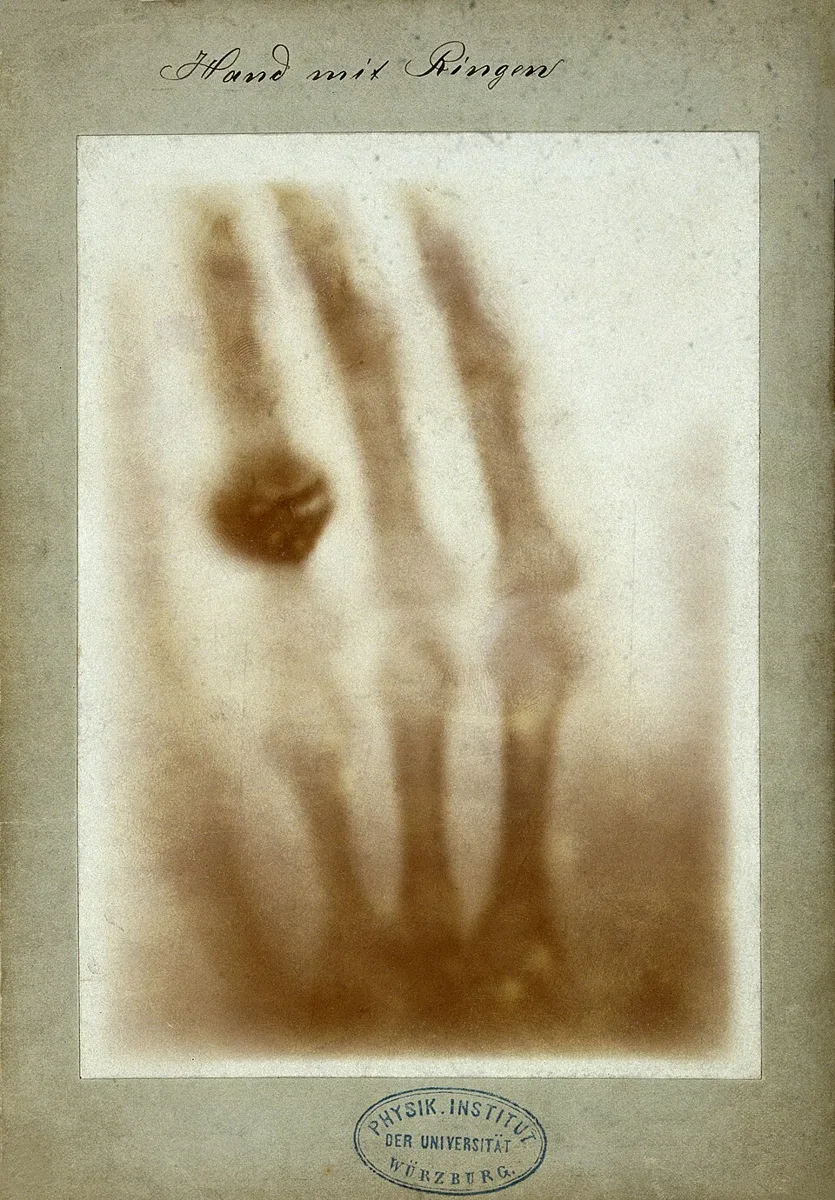
Historic X-Ray of Hand with Ring. This historic image displays the skeletal structure of a hand, complete with a prominent silhouette of a ring on one finger.Wilhelm Röntgen · Public domain · via Wikimedia CommonsView source ↗
Prompt
Transform this image into an X-Ray Radiograph with luminous white bones or dense internal structures, translucent charcoal soft tissues, and a nearly black background. Use stark monochrome contrast, ghostly overlapping silhouettes, faint photographic plate grain, and softly glowing structural edges. Keep the original subject and composition.
Wilhelm Röntgen: Hand mit Ringen (Hand with Rings), 1895.
Try it out
Hold your hand over abright flashlight in adark room to see how light passes through flesh but not bone.
Background
In late 1895, physicist Wilhelm Röntgen was experimenting with acathode-ray tube3in adarkened room. He noticed anearby fluorescent screen glowing. He discovered anew kind of invisible ray was escaping the tube. These "X-rays" could pass through cardboard, wood, and human flesh. He soon placed his wife Anna Bertha's hand between the tube and aphotographic plate. After development, the plate revealed her bones and wedding ring. This subtractive shadow-casting proved that different materials absorb radiation at different rates. The discovery revolutionized medicine and physics immediately.
Distinction
Unlike astandard photograph that captures reflected visible light, aradiograph captures ashadow map of interior density. Surface colors and textures remain invisible.
Aftermath
It teaches that human vision captures only atiny fraction of the electromagnetic spectrum.
Further examples · 11 images
02Printed Radiograph of a Mummified HandA printed reproduction reveals the internal bone structure of a mummified hand, surrounded by the faint outline of its preserved flesh. It demonstrates the radiographic technique, as unabsorbed electromagnetic radiation passes through less dense tissues to expose the plate, leaving shadows where denser material blocked the path.William Henry Meadowcroft · Public domain · Wikimedia Commons03Vintage Radiograph Print of a HandThe bones of a hand and wrist are sharply defined as dark structures against a lighter background in this early print. It exemplifies the effect by capturing the differential absorption of rays, forming a high-contrast shadow image of dense matter on the photographic medium.Unknown author · CC BY 4.0 · Wikimedia Commons04Dental Radiograph of Teeth and JawboneThis dental image shows the roots of several teeth embedded in the jawbone, with fine metal instruments appearing completely opaque. It acts as a clear radiograph, using electromagnetic rays to penetrate the gum tissue while being absorbed by the dense enamel, bone, and metal to create structural shadows.Bin im Garten · CC BY-SA 3.0 · Wikimedia Commons05Dental X-Ray Showing a Crowned ToothA single tooth fitted with a dense, bright crown and a treated root canal is visible alongside neighboring teeth within the jaw. This illustrates the radiographic process, showing how solid artificial materials and dense organic bone absorb radiation to form stark, high-contrast silhouettes against the exposed softer areas.Bin im Garten · CC BY-SA 3.0 · Wikimedia Commons06Close-Up Dental Radiograph with CrownAn extreme close-up of a jawbone reveals a tooth topped with an opaque artificial crown and a bright line indicating a root canal. The visual perfectly captures the radiographic effect, where dense dental materials and bone block electromagnetic rays from exposing the film, creating distinct, ghostly shadows.Bin im Garten · CC BY-SA 3.0 · Wikimedia Commons07Blue-Tinted Radiograph of a Human HandPresented in a blue tint with a measurement scale, this image clearly outlines the articulated bones of a human hand. It serves as a striking radiograph, highlighting how X-rays expose the background through soft skin and muscle while being absorbed by the skeletal structure to yield a high-contrast shadow.SarahCartwright · CC BY-SA 2.0 · Wikimedia Commons08Modern Blue X-Ray of a HandA human hand is displayed with its internal skeletal anatomy rendered as bright, solid shapes against a deep blue background. The technique is evident in the way denser bone tissue obstructs the invisible rays, leaving unexposed shadows that reveal the internal structure in high contrast.SarahCartwright · CC BY-SA 2.0 · Wikimedia Commons09Historical Positive Print of Hand X-RayA historical photographic print displays the skeletal structure of a hand, with a dark ring encircling one finger. This is an early X-ray radiograph printed as a positive, where dense objects like bones and metal cast shadows that are inverted into dark shapes.Arthur Schuster (1851–1934) · CC BY 4.0 · Wikimedia Commons10Modern Chest X-Ray RadiographA medical chest scan reveals the rib cage and spine as bright structures framing dark, transparent lungs. It is a standard X-ray radiograph where dense bone matter absorbs electromagnetic rays, leaving unexposed white areas on the negative film.Mahdi2ha · CC BY-SA 4.0 · Wikimedia Commons11Vintage Positive Print X-Ray of Two HandsA vintage photographic print shows the articulated bones of two adjoining hands as dark silhouettes against a lighter background. The image demonstrates the radiographic process by revealing the internal dense structures that blocked the invisible rays from exposing the original photographic plate.Unknown author · CC BY 4.0 · Wikimedia Commons
Question
Why do bones appear white on early X-ray plates?
Show possible answer +Hide answer −
Bones absorb the X-rays, blocking them from hitting the photographic plate. The unexposed areas become white after chemical development.