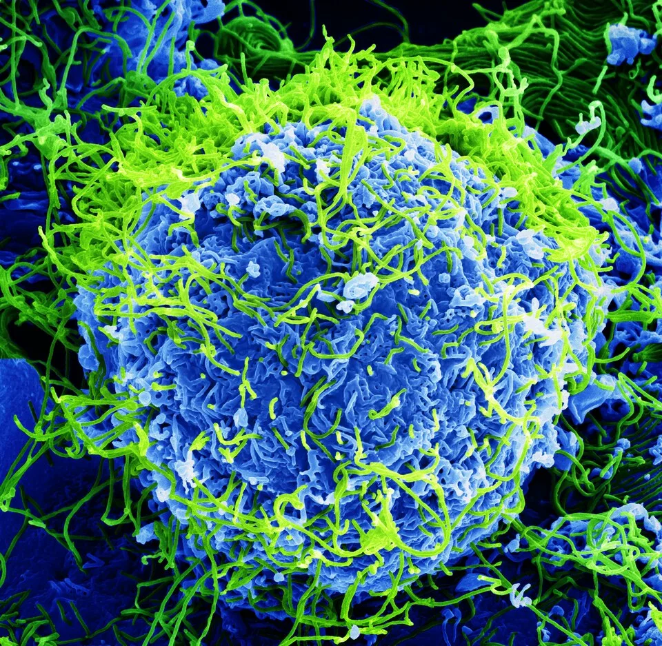
Filaments Across a Cellular Surface. Bright green filaments weave across a blue, deeply folded cellular surface.BernbaumJG · CC BY 4.0 · via Wikimedia CommonsView source ↗
Prompt
Transform this image into a Scanning Electron Micrograph, rendering surfaces in sculptural grayscale with exceptionally deep focus, crisp microscopic textures, bright raised edges, and dark crevices. Add fine detector noise and directional relief shading, making familiar materials resemble magnified specimens. Keep the original subject and composition.
Grey surfaces show [deep focus], crisp textures, and asculptural, three-dimensional appearance. [Bright edges]often stand against dark recesses; false colour may highlight selected structures.
Principle
Afocused electron beam scans the specimen, while adetector measures signals such as [secondary electrons1]emitted near its surface. Signal strength becomes image brightness; the beam’s narrow convergence angle provides alarge depth of field2.
Compare an SEM image of pollen with alight micrograph, noting which raised and recessed features remain sharp.
Background
Scanning electron microscopy developed before the 1960s, but commercial instruments helped bring it into wider laboratory use. Cambridge Instruments introduced its Stereoscan in 1965. Unlike acamera, an SEM builds an image by scanning an electron beam across aspecimen. Secondary electrons reveal surface detail, while detector position influences apparent shading. Conventional preparation often includes drying biological specimens and applying athin conductive coating3. These steps can alter delicate structures. The recorded signal has no inherent visible colour. Colour added afterward can separate structures or guide interpretation.
Distinction
Unlike transmission electron microscopy, which commonly reveals internal structure through thin specimens, SEM usually emphasizes surfaces. Its sculptural shading suggests depth, but asingle image is not athree-dimensional measurement.
Aftermath
SEM images teach us that convincing depth and detail can arise from measured electron signals rather than reflected light.
Further examples · 11 images
02Paired Microbes on a Folded SurfaceTwo yellow, oval microbial forms rest against a grey-green surface of folds and hollows. Finely resolved surface textures, bright rims, and dark recesses display the sculptural character of a false-coloured scanning electron micrograph.NIAID · CC BY 2.0 · Wikimedia Commons03Golden Bacterial ClustersGolden spherical bacteria gather in clusters across a green substrate, above an instrument readout and micrometre scale. Granular surface detail and the secondary-electron detector notation identify a false-coloured scanning electron micrograph.Janice Haney Carr, Centers for Disease Control and Prevention · Public domain · Wikimedia Commons04Purple Bacterial ColoniesDense clusters of purple spherical bacteria spread across an olive-coloured surface, with a ten-micrometre scale below. Resolved rounded contours and the secondary-electron acquisition readout demonstrate scanning electron microscopy with added colour.CDC / Jeff Hageman, M.H.S. / Janice Haney Carr · Public domain · Wikimedia Commons05Orange Bacteria on a Blue SubstrateOrange spherical bacteria form scattered chains and clusters on a blue, subtly ridged surface. Crisp surface relief, a micrometre scale, and secondary-electron imaging metadata establish this as a false-coloured scanning electron micrograph.CDC / Jeff Hageman, M.H.S. / Janice Haney Carr · Public domain · Wikimedia Commons06Golden Rods Within Blue Surface FoldsTwo golden rod-shaped forms lie within folded blue structures amid numerous small rounded bodies. Bright fold edges, dark cavities, and sharply rendered surface textures show scanning electron microscopy enhanced with selective false colour.NIAID · CC BY 2.0 · Wikimedia Commons07Yellow Microbes Among Red FoldsYellow spherical microbes cluster around broad red folds with finely wrinkled surfaces. The strong three-dimensional relief and detailed textures across multiple depths exemplify false-coloured scanning electron imaging.NIAID_Flickr · CC BY 2.0 · Wikimedia Commons08Segmented Surface in Electron ReliefA grey segmented specimen displays overlapping ridges, fine surface wrinkles, and sparse projecting hairs above a 200-micrometre scale. Bright ridge edges against dark recesses and extensive sharply resolved relief are characteristic of scanning electron microscopy.Hannah G Watson, Andrew T Ashchi, Glen S Marrs, Cecil J Saunders · CC BY 4.0 · Wikimedia Commons09False-Coloured Cell with Surface ParticlesA rounded blue cell is covered with curling green surface folds and numerous small yellow particles. False colouring highlights structures whose crisp textures, luminous edges, and deep recesses reveal the sculptural surface detail of scanning electron microscopy.NIAID · CC BY 2.0 · Wikimedia Commons10Yellow Spheres on a Folded Cell SurfaceFour yellow spherical bodies rest along the crest of a folded surface coloured blue and brown. Scanning electron microscopy renders their granular skins and the surrounding membrane relief with pronounced depth, while added colour separates the structures.NIAID · CC BY 2.0 · Wikimedia Commons11Perforated Microscopic Shells and PlatesThree monochrome panels show perforated microscopic shells and clusters of ringlike plates, accompanied by micrometre scale bars. Bright rims, dark openings, and sharply resolved surface ornament demonstrate the topographic detail characteristic of scanning electron micrographs.Mathias Haunost, Ulf Riebesell, Francesco D’Amore, Ole Kelting and Lennart T. Bach · CC BY-SA 4.0 · Wikimedia Commons
Question
Does a coloured SEM image show the specimen’s natural visible colours?
Show possible answer +Hide answer −
No. The electron signal does not record visible colour; added colours usually distinguish structures or support interpretation.