1840s, early work by Alfred Donné and William Henry Fox Talbot
Typeface · IM Fell French Canon
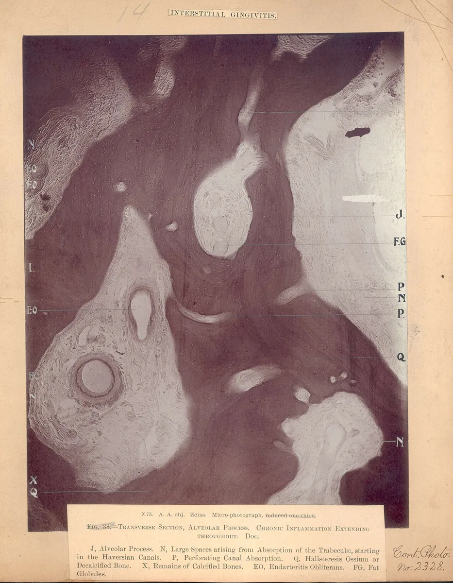
Historical Tissue Section Photomicrograph. An enlarged tissue section fills a historical plate with branching dark masses, pale spaces, and anatomical reference letters.National Museum of Health and Medicine · CC BY 2.0 · via Wikimedia CommonsView source ↗
Prompt
Apply a Photomicrography treatment that makes existing surfaces resemble magnified translucent specimens, revealing fine cellular meshes, granular inclusions, and delicate internal striations. Use transmitted illumination, shallow focus, restrained monochrome tones, and a softly darkened circular field without cropping essential details. Keep the original subject and composition.
Tiny structures fill the frame: [enlarged cells], insect scales, or mineral crystals reveal [fine internal patterns]. Some images retain acircular field with dark edges.
Principle
Amicroscope forms a[magnified optical image], which exposes alight-sensitive surface or reaches an electronic sensor. Illumination, focus, and the resolving power1of the optics determine which details become visible.
Example
Alfred Donné and Léon Foucault: Atlas du cours de microscopie, 1845.
Try it out
Photograph aprepared plant slide through amicroscope eyepiece, then compare visible cell boundaries at two magnifications.
Background
During the 1840s, experimenters joined photography to microscopy to record structures too small for unaided sight. Donné and Foucault made daguerreotypes2through microscopes. Their 1845 atlas presented engraved reproductions based on these photographs, rather than photographs pasted into the book. Talbot also explored photographing microscopic subjects during photography’s early years. The microscope supplied the enlarged optical image; photographic exposure recorded it on asensitive surface. Development and fixing3made the record visible and stable. Careful lighting mattered because magnification alone could not reveal every structure.
Distinction
Photomicrography records subjects through amicroscope; macro photography generally records small subjects with acamera lens alone. Microphotography instead makes extremely small photographs, such as microfilm records.
Aftermath
Photomicrography shows that greater image size does not automatically mean greater resolved detail.
Further examples · 11 images
02Crystalline Patterns in Polarized LightSweeping bands of blue, violet, green, and gold reveal finely striated crystalline structures. Magnification and polarized illumination expose crystal patterns and optical colors characteristic of polarized-light photomicrography.Janeklass · CC BY 4.0 · Wikimedia Commons03Foucault’s Daguerreotype PhotomicrographsA historical display assembles circular microscope photographs attributed to Léon Foucault, showing blood corpuscles and other minute specimens. The visible specimen images document early daguerreotype photomicrography rather than merely describing the technique.Unknown author · CC BY 4.0 · Wikimedia Commons04Ice Crystal With Radial ChannelsA pale ice crystal displays radial, club-shaped channels within a dark circular field on a mounted photographic print. Microscope enlargement makes the crystal’s delicate internal growth patterns visible, clearly demonstrating photomicrography.Wilson Bentley · Public domain · Wikimedia Commons05Hexagonal Ice PlateA hexagonal ice plate shows six triangular sectors, scalloped internal margins, and a small central feature against a dark field. The photograph records fine crystal structure at microscopic scale, exemplifying photomicrography of ice.Wilson Bentley · Public domain · Wikimedia Commons06Sixfold Ice Crystal Growth PatternsA sixfold ice crystal carries nested angular outlines and branching channels around a flower-shaped center. Its magnified photographic image reveals subtle growth boundaries that make it a clear example of photomicrography.Wilson Bentley · Public domain · Wikimedia Commons07Petal-Lobed Ice CrystalSix broad, petal-like ice lobes radiate from a central point, with fine needles and trapped markings visible inside them. The microscope photograph exposes small-scale crystal details within a dark circular field.Wilson Bentley · Public domain · Wikimedia Commons08Ice Crystal with Patterned CenterA six-armed ice crystal displays rounded projections and a finely patterned center against a dark circular field. This photographic print shows microscope magnification revealing the crystal’s delicate internal structure.Wilson Bentley · Public domain · Wikimedia Commons09Broad-Armed Snow CrystalA snow crystal has six broad, angular arms surrounding a small star-shaped center. Photomicrography enlarges the crystal to reveal fine branching marks and layered outlines within its arms.Wilson Bentley · Public domain · Wikimedia Commons10Branching Snow CrystalA snow crystal spreads six pointed branches, each edged with smaller, fernlike offshoots. The photomicrograph resolves intricate crystalline growth within a dark circular field.Wilson Bentley · Public domain · Wikimedia Commons11Banded Six-Pointed Ice CrystalA sharply pointed, six-armed ice crystal contains alternating light and dark bands and small rounded markings. Microscope photography makes these fine internal patterns visible across the enlarged crystal.Wilson Bentley · Public domain · Wikimedia Commons
Question
Why can two photographs at the same magnification show different amounts of detail?
Show possible answer +Hide answer −
Their optics may have different resolving power, and their focus or illumination may differ. Magnification enlarges the image but cannot restore missing detail.